
Hypocalcemia: A Nurse's Guide to Low Calcium Levels

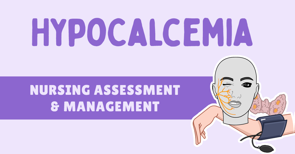
Hypocalcemia — low calcium in the blood — is an electrolyte abnormality you can't afford to miss. Calcium drives muscle contraction, nerve transmission, blood clotting, and the electrical activity of the heart, so when levels drop, the consequences can range from tingling fingertips to seizures and lethal arrhythmias.
Nutritional deficiencies, medications, kidney disease, and parathyroid problems can all pull calcium down. As the nurse at the bedside, knowing how to recognize the signs, interpret the labs, and act on them is what keeps these patients safe.
This guide covers why calcium matters, how the body regulates it, the lab values you need to know, the causes and symptoms of hypocalcemia, and exactly how it's assessed, treated, and monitored — including the cardiac arrhythmias low calcium can trigger.
Why Is Calcium Important in the Body?
Calcium is far more than a bone mineral. It's an essential electrolyte that keeps multiple systems running, which is exactly why a low level can cause such varied — and sometimes dangerous — symptoms.


How Is Calcium Regulated?
Calcium is kept within a tight range by a feedback system involving three hormones acting on the bones, intestines, and kidneys. Understanding this loop explains nearly every cause of hypocalcemia.


Normal Calcium Levels & Hypocalcemia
Total serum calcium is what you'll see on a BMP or CMP. Here are the numbers to commit to memory:
What Are Ionized Calcium Levels?
When calcium is low, you'll often see an ionized calcium ordered. Here's why: ionized calcium is the physiologically active form. Total calcium includes both ionized and protein-bound calcium, and it's influenced by pH and albumin — so ionized calcium is the more accurate reflection of true calcium status.
Albumin & calcium
Albumin binds calcium, so a low albumin can make total calcium look falsely low. Correction calculators exist but aren't always accurate — if calcium and albumin are both low, check an ionized calcium to confirm the real status.
Causes of Hypocalcemia
Most causes trace back to a problem with PTH, vitamin D, the kidney, or binding of calcium out of the bloodstream.


The hyperventilation connection
Anxiety and hyperventilation blow off CO₂, causing respiratory alkalosis. Alkalosis makes calcium bind more tightly to albumin, dropping ionized calcium — which is why panic-attack patients get peri-oral numbness, tingling, and spasms. Having them breathe into a non-rebreather (not hooked to oxygen) helps them retain CO₂ and ease symptoms.
Nursing Assessment of Hypocalcemia
The hallmark of hypocalcemia is tetany — hyper-irritability of the peripheral nerves and the muscles they control. It ranges from mild tingling to dangerous spasms, and typically doesn't appear until calcium drops below about 7–7.5 mg/dL.


Vital signs may show an irregular heart rhythm (arrhythmia) and a decreased blood pressure. Otherwise, watch for weakness and painful muscles. For more on the neuro exam, see the cranial nerve assessment guide.

Treatment of Hypocalcemia
When a low calcium comes back, your first job as the nurse is a quick, structured response before treatment is even ordered:
Check for symptoms and confirm stability — including recent vital signs.
Place the patient on the cardiac monitor and get an ECG if not already done. Continuous monitoring is essential while infusing IV electrolytes.
Report the calcium level, your assessment, and the cardiac rhythm — flag any QT prolongation.
Have at least one IV; place a second line if hypocalcemia is severe.
Give the ordered calcium and supplements (below).
The specific treatment depends on severity and symptoms. Severe features that demand prompt IV repletion include tetany, arrhythmias, and seizures.

- Bolus: Calcium gluconate 1–2 g (90–180 mg elemental) in 50 mL D5W or NS over 10–20 min; may repeat. Raises calcium for only ~2–3 hours.
- Infusion: 10% calcium gluconate continuous drip — e.g., 11 g in 1000 mL NS/D5W at ~50 mL/hr.

- Calcium carbonate 1250 mg — 1 tab PO TID
- Calcium citrate 950 mg — 2 tabs PO BID

- Ergocalciferol (D2) or cholecalciferol (D3) 50,000 IU PO weekly × 6–8 weeks
- Calcitriol 0.25 mcg PO BID — preferred in severe hypocalcemia or kidney/liver disease (skips activation steps for faster correction)

Monitoring of Hypocalcemia
Close monitoring catches both under-correction and over-correction. Watch these parameters:


Hypocalcemia & Cardiac Arrhythmias
Calcium is central to cardiac conduction, so very low levels can produce distinct ECG changes and dangerous rhythms.

Arrhythmias associated with hypocalcemia include Torsades de Pointes (polymorphic VT that twists around the baseline), atrial fibrillation, ventricular tachycardia, PVCs, and AV blocks.
Torsades is an emergency
Torsades de Pointes can rapidly degenerate into ventricular fibrillation if not treated immediately. It's less common with hypocalcemia than with hypomagnesemia — but when QT is prolonged, stay vigilant.
Clinical Pearls

Master the rhythms low calcium causes
My video course ECG Rhythm Master, built for nurses, takes you deep into arrhythmias like Torsades, VT, and AFib. You'll be able to:
- Identify every cardiac rhythm with confidence
- Understand the pathophysiology behind each one
- Manage arrhythmias like an expert nurse
- Run emergency procedures — pacing, defibrillation, cardioversion
Related Guides


References
- Goltzman D. Clinical manifestations of hypocalcemia. UpToDate.
- Goltzman D. Diagnostic approach to hypocalcemia. UpToDate.
- Goltzman D. Etiology of hypocalcemia in adults. UpToDate.
- Goltzman D. Treatment of hypocalcemia. UpToDate.
- Schafer AL, Shoback DM. Hypocalcemia: Diagnosis and Treatment. Endotext / NCBI Bookshelf.
- Hypocalcemia: A clinical review. National Library of Medicine (PMC).
- Harding MM, et al. Lewis's Medical-Surgical Nursing: Assessment and Management of Clinical Problems. 11th ed. Elsevier.
Hello, I appreciate your writing very much. Could we please discuss your article on AOL further? I am in need of a specialist in this field to resolve my problem. Perhaps you are that individual. I look forward to seeing you.